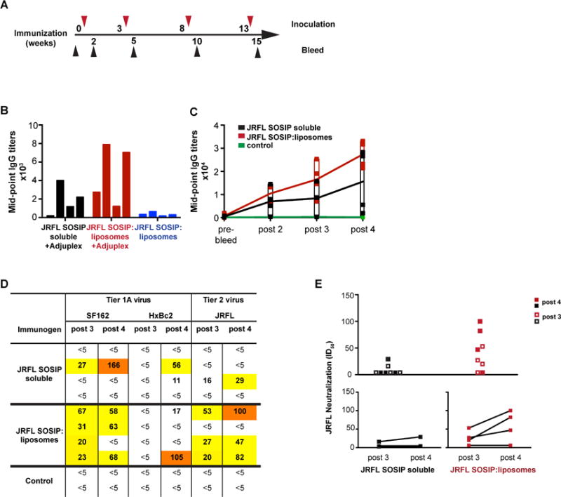
Figure 7. Immunogenicity of the JRFL SOSIP trimer-conjugated liposomes.

(A) Timeline of inoculations and bleeds. Bleeds were collected 2 weeks after each injection. (B) Mid-point IgG titers of individual rabbits immunized 3 times with 25 ug JRFL SOSIP protein as soluble trimers or conjugated to liposomes in the presence or absence of exogenous adjuvant Adjuplex were determined. Sera were collected 2 weeks after the 3rd injection and were analyzed by ELISA with JRFL SOSIP trimers captured on ELISA plate via the C-terminal His6tag. (C) Mid-point IgG titers of rabbits immunized 4 times with JRFL SOSIP soluble protein or JRFL SOSIP conjugated to liposomes. ELISA plates were coated with anti-His monoclonal antibody to capture JRFL SOSIP trimers via the C-terminal His6-tag. (D) Neutralization ID50 values of SF162, HxBc2, and JRFL viruses by antisera following the third and fourth inoculations. Control animals were inoculated with blank liposomes in Adjuplex. (E) Combined JRFL neutralization ID50 values elicited by the soluble trimers compared to the trimer-conjugated liposomes are plotted after third and fourth inoculations. Lower panel shows boosts in ID50 values after the fourth inoculation for both group of rabbits. See also Figure S5.
