Fig. 2.
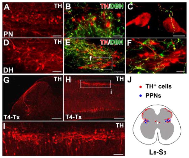
The distribution of TH+ cells in the rat lumbosacral spinal cord. A–F, In horizontal sections of the intact rat spinal cord, many TH+ cells are found in the lateral parasympathetic nuclei (PN) (A), lamina X, and superficial dorsal horn (DH) (D). Though a large number of descending fibers are TH+ and DBH+, lumbosacral TH+ cells and their projections are DBH− (B, C, E, F), indicating that they are not noradrenergic/adrenergic. G–I, Three weeks after T4–Tx, spinal TH+ cells and their processes remain in coronal (G) or longitudinal (H, I) sections of lumbosacral cord. Numerous TH+ cells are prominent in the parasympathetic regions of gray matter (H, I). TH+ axons extend segmentally and do not form long ascending projections. J, A schematic diagram illustrates the distribution of TH+ cells (red) in L6–S3 spinal segments in a transverse view of spinal cord. These subpopulations are connected via fibers in lamina I. Notably, TH+ cells are intermingled with parasympathetic preganglionic neurons (PPNs, blue) in the lateral autonomic regions. F and I are higher magnifications of regions boxed in E and H. Scale bars: A, D, 75 μm; B, 50 μm; C, 10 μm; E, I, 100 μm; F, 30 μm; G, 200 μm; H, 400 μm. (For interpretation of the references to color in this figure legend, the reader is referred to the web version of this article.)

