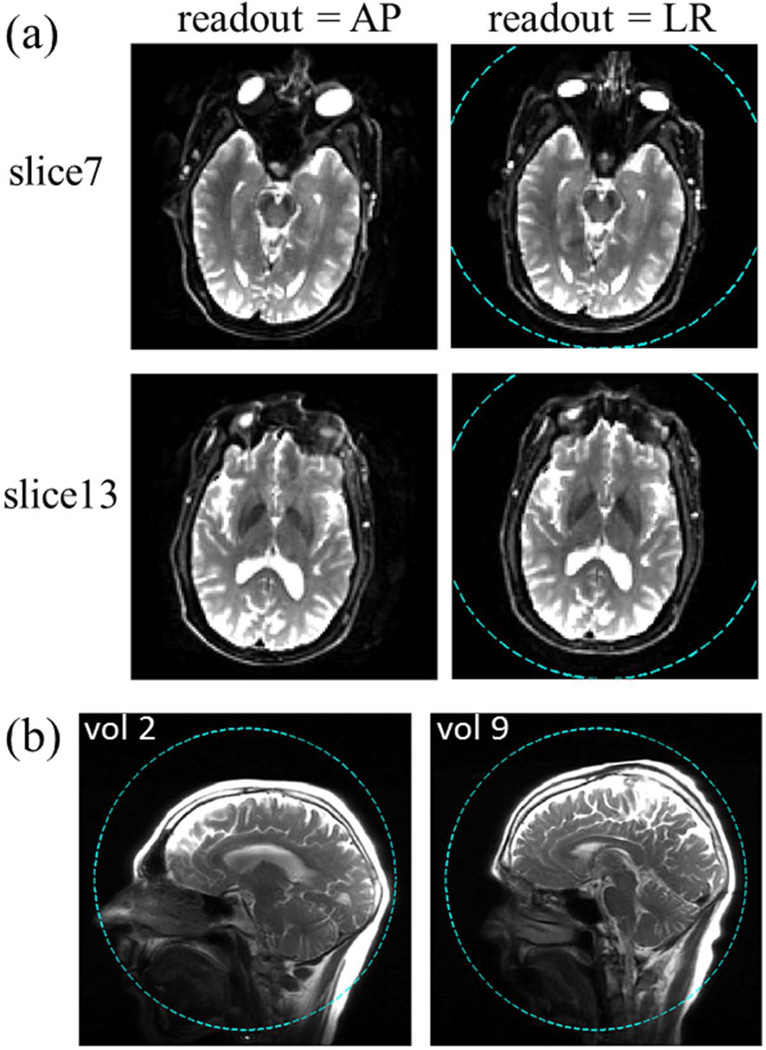
Figure 7.
(a) Axial EPI images of a volunteer with different readout directions. Two representative slices are shown with thickness = 3.2 mm, acquired with a slew rate of 500 T/m/s and gradient amplitude of 46 mT/m in the readout portion of the sequence. Note the relatively mild distortion around the sinus area compared to typical single-shot EPI images acquired with standard whole-body gradients. (b) Sagittal localizer images of two volunteers with a short (left) and a long (right) neck. In both (a) and (b), dashed-line circle shows the 26 cm DSV linearity region.

