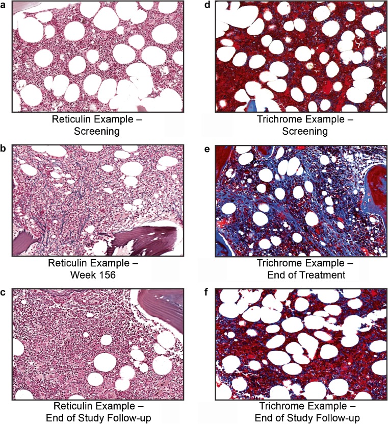
Fig. 2.
Images from bone marrow biopsies of patients with ITP treated with romiplostim. Bone marrow images of a patient with reversible increased reticulin (silver stain) (a–c) and of another patient with reversible collagen (trichrome stain) (d–f). Images are arranged as follows: screening (a, d), end of treatment (b, e), and follow-up after discontinuation (c, f). As not all biopsies were stained at the same time; there were slight differences in staining intensity, brightness, and tone between timepoints for a single patient. To more easily allow for comparison of images of a single patient and to correct for scanning defects, brightness, contrast, and saturation were adjusted using PowerPoint. ITP immune thrombocytopenia

