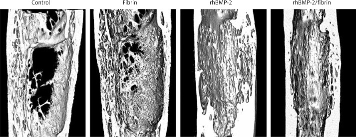
Figure 4.
The 3D micro-CT images 8 weeks after drug treatment revealed a superior quality of ossification of the distracted gap in the rabbits treated with rhBMP-2 + fibrin compared to others. Cross-sectional images at the center of the lengthened segment indicated that the rhBMP-2 + fibrin group showed the thickest cortical bone

