
Figure 2.
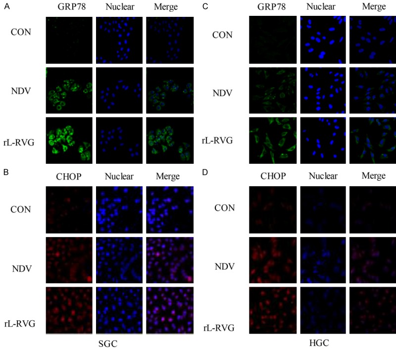
Expression of ER stress-related proteins in infected SGC and HGC cells. The nuclei were stained with Hoechst 33342. The cells had been infected with virus for 24 h. ER stress-related protein expression was monitored by immunofluorescence microscopy (x200 magnification). A and B. For SGC, C and D. For HGC. A and C. GRP78 protein (green) was significantly more strongly expressed in the NDV and rL-RVG groups, and the rL-RVG-infected group exhibited the strongest expression. Similarly, in B and D. CHOP protein expression was strongest in the rL-RVG-infected group.