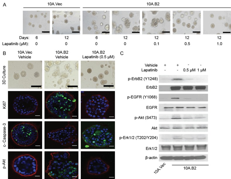
Figure 2.

Lapatinib inhibited PI3K-Akt pathway and prevented the transition of 10A.B2 cell acini from atypia to DCIS in 3D model. A. Phase contrast microscopy images of 10A.Vec and 10A.B2 cells treated with Vehicle or Lapatinib at indicated concentrations in 3D culture. Scale bar represents 200 µm. B. Immunofluorescence staining analysis of Ki-67, cleaved caspase-3 and p-Akt in 10A.Vec, 10A.B2 and 10A.B2 cells treated with lapatinib (0.5 µM) for prevention study in 3D culture on day 12. Ki-67, cleaved caspase-3, and p-Akt are shown in green. Laminin V is shown in red. DAPI is shown in blue. Scale bar represents 50 µm. Light microscopy photos (top panel) were used as reference. Scale bar represents 200 µm in light microscopy images. C. Western blotting analysis of p-ErbB2, p-EGFR, p-Akt and p-Erk1/2 expression in protein level in indicated cells with Vehicle or Lapatinib treatment.
