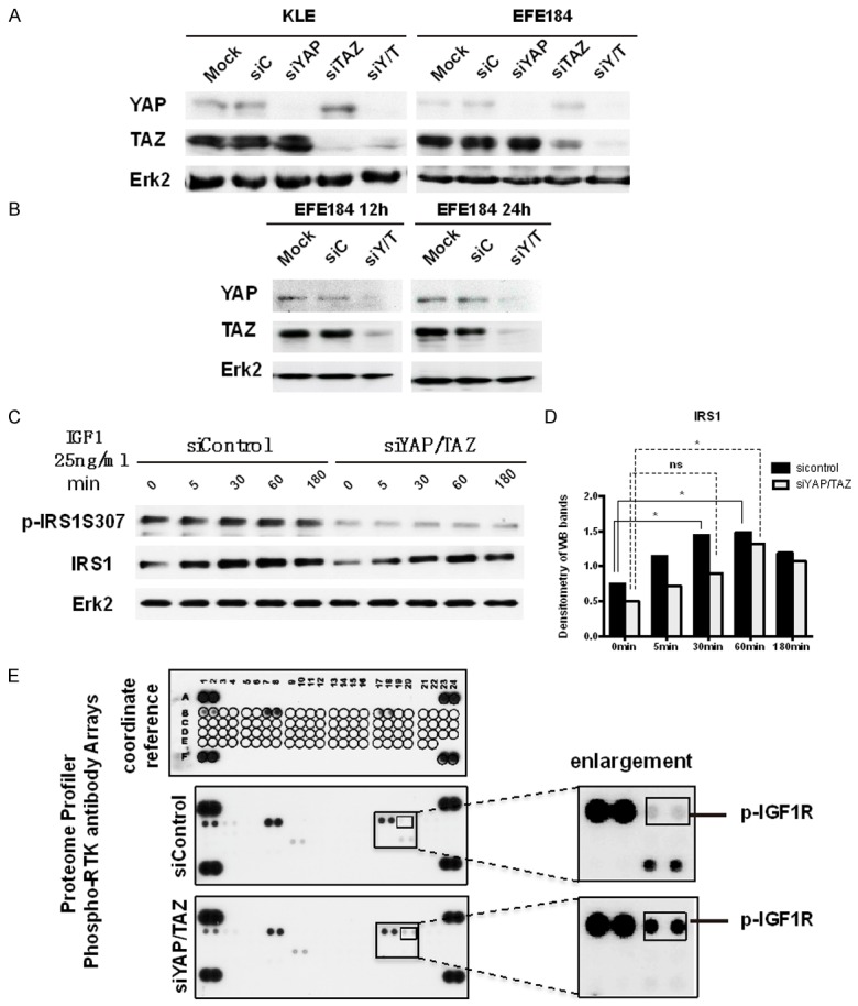
Figure 4.

siYAP/TAZ decreased the phosphorylation of IRS1 but increased the phosphorylation of IGFR1. A. KLE cells and EFE184 cells were transfected with mock, siControl, siYAP, siTAZ and siYAP/TAZ (siY/T). At 48 h after transfection, cells were collected for Western blot with the indicated antibodies. Erk2 was used as a loading control. B. EFE184 cells were transfected with mock, siControl, siY/T. At 12 h and 24 h after transfection, cells were collected for Western blot with the indicated antibodies. Erk2 was used as a loading control. C. In KLE cells, YAP and TAZ were knocked down by siRNA, after 24 h, IGF1 25 ng/ml was administrated to cells for 5 min, 30 min, 60 min, 180 min. And then, cells were collected and processed to western blotting with according antibodies. It shows siYAP/TAZ decreased the IRS1 expression level. D. The bar graph is the densitometry of figure c, which indicates siYAP/TAZ decreased the IRS1 expression level significantly. Statistical significance was calculated using one way ANOVA and Dunnett’s multiple comparisons test compared with 0 min. *P < 0.5, ns: non-significant. E. KLE cells were transfected with siYAP/TAZ. After 24 h, cells were processed to Proteome profiler phosphor-RTK antibody arrays (R&D Systems Catalog Number ARY001B). Every antibody was repeated as two dots. siYAP/TAZ increased the p-IGF1R expression, which is shown in two smallest frames.
