Figure 2.
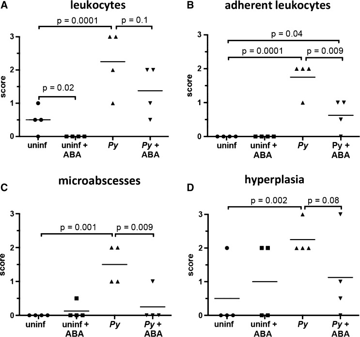
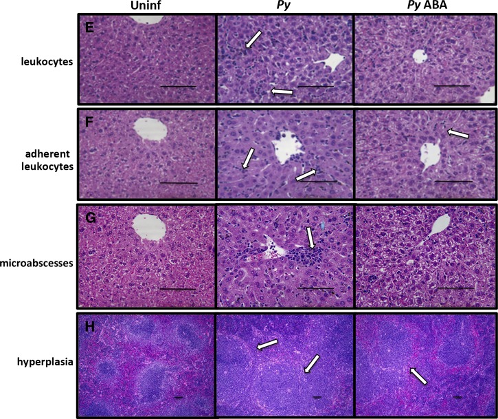
Oral supplementation of abscisic acid (ABA) decreased tissue inflammatory responses of Plasmodium yoelii infected mice. (A) The livers of P. yoelii infected mice had significantly more nonadherent sinusoidal leukocytes than uninfected controls. ABA reduced the hepatic nonadherent leukocytes of uninfected mice, but failed to significantly reduce the hepatic nonadherent leukocytes of infected mice. (B) The livers of P. yoelii infected mice again had significantly more adherent sinusoidal leukocytes than uninfected controls. ABA had no effect on the hepatic adherent leukocytes of uninfected mice, but highly significantly reduced the hepatic adherent leukocytes of P. yoelii infected mice. (C) The livers of P. yoelii infected mice had significantly more microabscesses than uninfected controls. ABA had no effect on the hepatic microabscesses of uninfected mice, but highly significantly reduced the hepatic microabscesses of P. yoelii infected mice. (D) The spleens of P. yoelii infected mice had significantly more lymphoid hyperplasia of periarteriolar lymphoid sheaths than uninfected controls. ABA had no effect on the splenic lymphoid hyperplasia of uninfected mice, but showed a trend, though not statistically significant, toward reducing splenic lymphoid hyperplasia of P. yoelii infected mice. (E) Liver sections with arrows indicating sinusoidal nonadherent leukocytes. (F) Liver sections with arrows indicating adherent sinusoidal leukocytes. (G) Liver sections with a sinusoidal microabscess indicated by the arrow. (H) Spleen sections with lymphoidal hyperplasia of the periarteriolar lymphoid sheaths indicated by arrows. Bars = 100 μm; Py = P. yoelii-infected; Uninf = uninfected.


