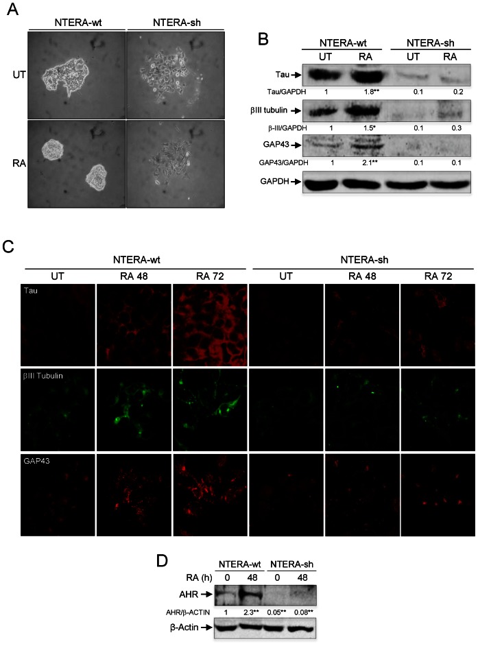
Figure 1.
AHR knockdown impairs RA-induced differentiation. (A) NTERA-wt and AHR knockdown NTERA-sh cells were grown in 2D cultures and clone formation analyzed 48 h later in absence (UT) or presence of 1 μM RA. (B) Both cell lines were left untreated (UT) or treated with 1 μM RA for 48 h. Total cell extracts were analyzed for the expression of neuronal markers Tau, βIII-tubulin and GAP43 by immunoblotting. GAPDH was used to normalize protein expression. (C) Tau, βIII-tubulin and GAP43 were also analyzed by immunofluorescence in untreated (UT) or RA-treated NTERA-wt and NTERA-sh cells using a Fluoview FV1000 confocal microscope. (D) NTERA-wt and NTERA-sh cells were left untreated or treated with 1 μM RA for 72 h and total AHR protein levels determined by immunoblotting. β-Actin was used as normalization control. Panels A–D: n = 4 biological replicates. *P < 0.05 and **P < 0.01. Data are shown as mean ± SD.

