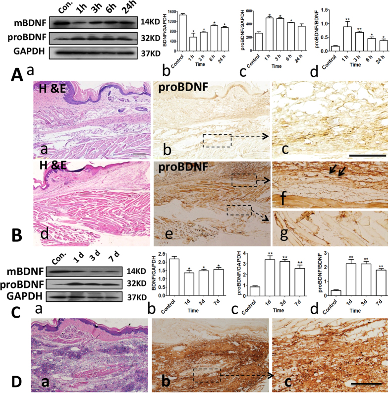
Figure 1. Upregulation of proBDNF in the local tissue in acute and persistent inflammatory pain in mice.
(A) Representative Western blot (a) and their semi-quantitative analyses of mature BDNF (b), proBDNF (c) and their ratio (d) in the local tissue after 10 μL 5% formalin intra-plantar injection into Kunming mice (*p < 0.05, **p < 0.01 versus control, one-way ANOVA followed by Dunnett’s Multiple Comparison post hoc test, n = 3 per group). (B) H&E staining (a and d) and immunohistochemsitry (b,c and e–g) of proBDNF in the foot skin at 3 h post-formalin injection. proBDNF is expressed in the epidermis, basal layer and subcutaneous layers in the foot skin (b,c); Higher magnification (box in b) showing proBDNF is also mildly expressed in the nerve fibers in the control plantar (c); Responding to peripheral inflammation by 5% formalin intra-plantar injection, intensive proBDNF immunoreactivity is observed and mainly localized in the inflammatory cells (f, black arrows) and nerve fiber-like structures (g). Scale bars: 50 μm, 3 replicates, n = 3 per group. (C) a, Representative Western blot of proBDNF and mBDNF; b–d, Semi-quantitative analyses of mBDNF, proBDNF and their ratio in the inflamed tissue after Complete Freund Adjuvant (CFA, 10 μL) intra-plantar injection into Kunming mice (*p < 0.05, **p < 0.01versus control, one-way ANOVA followed by Dunnett’s Multiple Comparison post hoc test, n = 3 per group). (D) Histological staining (a) and proBDNF immunohistochemistry (b,c) in the plantar at 1 day post-CFA injection; c, higher magnification of box in b showing that proBDNF is highly expressed in the inflammatory cells. Scale bar, 100 μm, 3 replicates, n = 3 per group. Data bars represent mean ± s.e.m.

