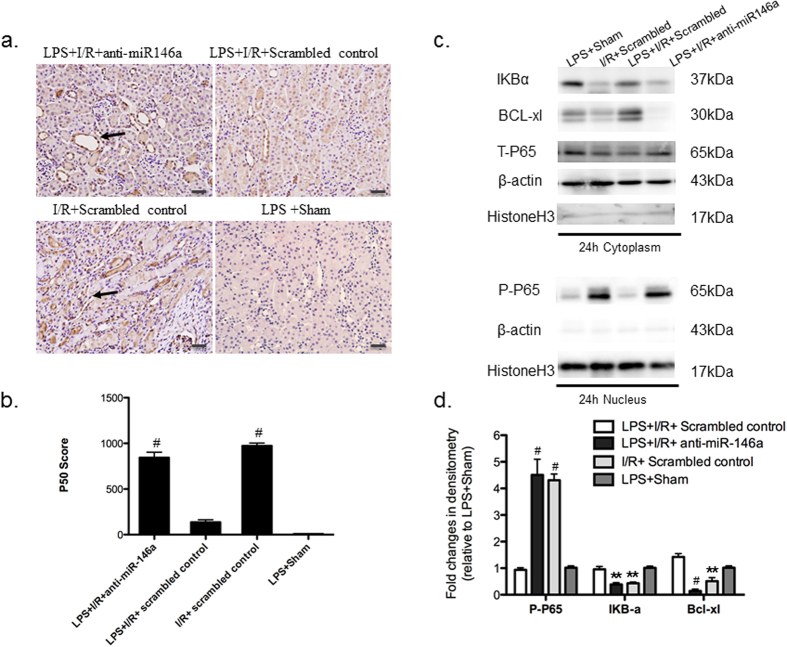
Figure 7. The link of miR-146a and NF-κB activation.
(a) Immunohistochemistry shows positive nuclear NF-κB p50 staining 24 h after I/R in mice exposed to LPS with or without miR-146a knockdown (original magnification x200, Bar = 50 um) (b) Nuclear NF-κB p50 expression was scored in mouse kidney. (n = 6 mice per group, #P < 0.001, vs. LPS + I/R + Scrambled control group). The specificity of the staining was further demonstrated by the absence of signals in LPS + Sham group (c) Kidney lysates of 24 h groups were probed with specific antibody against phosphorylated P65 (p-p65) (nuclear extracts), and IkBα, BcL-xL, T-P65 (cytosolic extracts). Co-detection of Histone H3 (nuclear) and β-actin (cytoplasm) were performed to assess equal loading. A significant increase in nuclear NF-κB p65 expression, cytosolic IkBα and BcL-xL degradation at 24 h was observed. (d) The Western blots from all experiments were quantified by densitometry analysis. The ratios of phosphor-protein to total protein for p65 were calculated. The fold changes relative to LPS + Sham protein are shown. (n = 3, #P < 0.001, **P < 0.01, vs. LPS + I/R + Scrambled control group).

