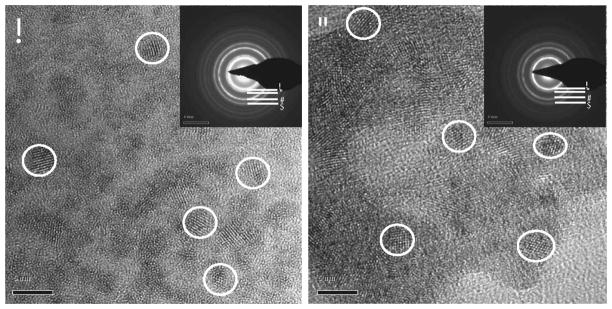
FIG. 1.
High-resolution transmission microscopy image of CNPs. Panel A: CNP-18 synthesized using wet chemical method [inset: selected area electron (SAED) pattern]. Panel B: CNP-ME synthesized using microemulsion process (inset: SAED pattern). Inset key: A, B, C and D represent 111, 200, 220 and 311crystal lattice planes, respectively, of CNP fluorite lattice structure. Scale bar = 5 nm.

