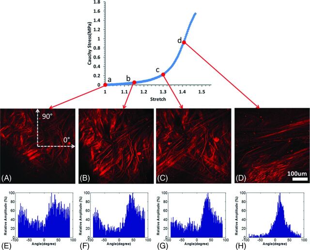
Fig 5.
Abluminal view of collagen fiber recruitment during uniaxial loading of aneurysm sample CA26 (green in Figs 1–3) obtained by using the uniaxial MPM system. The images were obtained at stretches of 1.0 (A), 1.15 (B), 1.3 (C), and 1.4 (D) and were formed from a projection of stacks over an approximately 95-μm depth of tissue. E–H, Histograms of fiber-orientation distribution of the MPM images at stretches of 1.0, 1.15, 1.3, and 1.4, respectively. The horizontal direction on the image is 0°, and the vertical direction is 90° (as shown in A).

