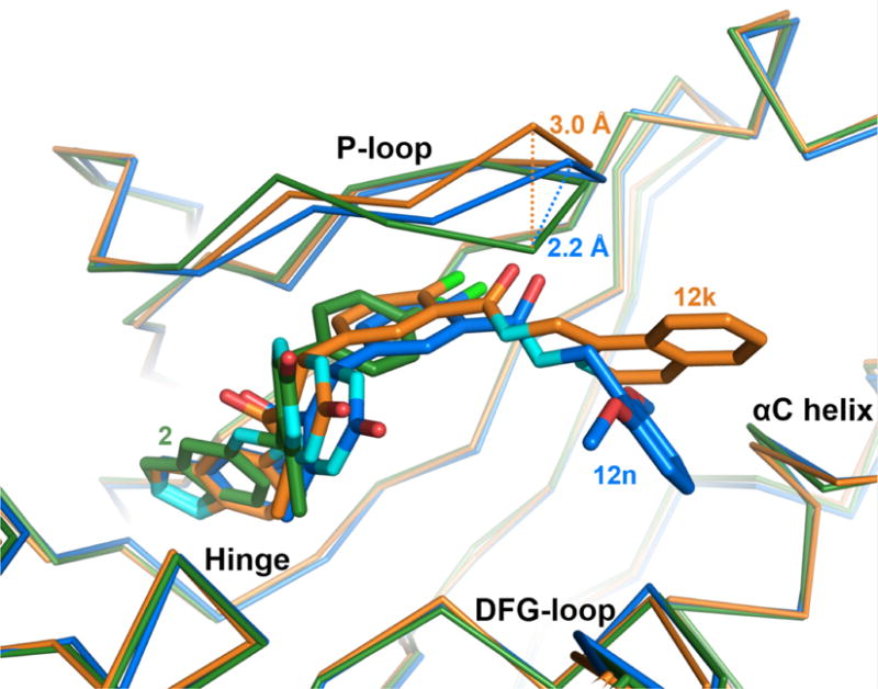
Figure 4.

Adaptive structural changes in the GRK2 P-loop. Compared to the P-loop conformation when bound to compound 2 (green), the Cα carbon of Gly201 shifts away from the binding site by 2.2 Å when bound to compound 12n (blue), 12h or 12r (not shown), and by 3.0 Å when bound to 12k (orange). The magnitude of the shift thus appears to depend on the size of the D-ring.
