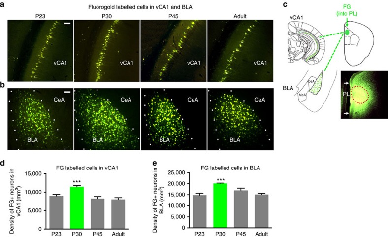
Figure 2. Dynamic developmental surge in neural connectivity to mPFC.
Representative images of FG-labelled neurons in (a) hippocampus ventral CA1 (vCA1) and (b) basolateral amygdala (BLA), Scale bars, 50 μm. (c) Schematic of FG injection in prelimbic cortex (PL) and FG+ cells throughout vCA1 and BLA. Outlined area denotes injection site, with surrounding diffusion maintained within PL boundaries. Quantification of density of FG (FG+) retrograde labelled neurons in showed a significant main effect of age in (d) vCA1, P<0.0005, F(3,16)=11.58, vCA1 (P23 8970.7±401.3; P30 11441.1±368.8; P45 8254.4±565.1; P90 8024.6±479.5) and a significant main effect of age in (e) BLA, P <0.0005, F(3, 16)=12.2, BLA (P23 14809.1±847.3; P30 20124.1±165.6; P45 16995.1±973.0; P90 15114.8±520.5).

