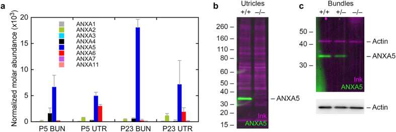
Figure 1. Protein chemistry analysis of annexins in vestibular hair cells.
(a) Annexin paralog expression in developing and mature mouse utricle and hair bundles using quantitative mass spectrometry. The y-axis indicates the relative fraction (concentration) of each annexin in the appropriate sample. Samples were P5 hair bundles (P5 BUN), P5 whole utricle (P5 UTR), P23 hair bundles (P23 BUN), and P23 whole utricle (P23 UTR). Four replicates each of 100 ear-equivalents (bundles) or 10 utricles were analyzed. Mean ± SD are plotted. (b) Protein immunoblotting of ANXA5 in P23 wild-type and Anxa5-mutant utricles. One utricle was analyzed for each genotype; the immunoblot was first stained with India ink (magenta) to visualize total protein, then was probed with anti-ANXA5 (green). The band of the correct molecular mass (~33 kD) seen in wild-type utricles was completely absent from the knockout utricles. (c) Protein immunoblotting of ANXA5 in P23 wild-type and Anxa5-mutant hair bundles (7 ear-equivalents per age). Genotypes indicated above lanes. Because of its abundance in bundles, actin was the only protein readily detected by the ink stain; ANXA5 was detected by immunoblotting in wild-type and heterozygous bundles, but not in homozygous knockout bundles. Actin was detected by immunoblotting at similar levels in all three lanes.

