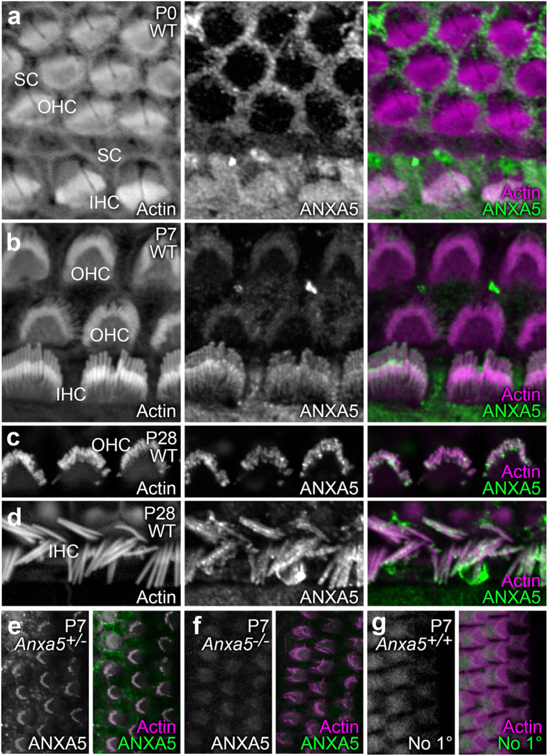
Figure 2. Localization of ANXA5 in organ of Corti stereocilia shifts during postnatal development.
For (a–e) samples were labeled with rhodamine-phalloidin (left panels and magenta in right-panel merges) and anti-ANXA5 (middle panels and green in right-panel merges). (a) At P0, inner hair cell (IHC) stereocilia had ANXA5 labeling throughout the bundle, whereas labeling was absent in outer hair cells (OHC). Supporting cell (SC) apical surfaces were strongly labeled. (b) At P7, ANXA5 was visible in OHC stereocilia and displays distinct localization to the tips, as well as periphery of IHC stereocilia. (c) At P28, ANXA5 as observed at stereocilia tips and shafts of OHCs. (d) ANXA5 stereocilia tips and shafts of IHCs at P28. (e–g) Labeling controls; antibody channel alone is in the left panel, and the merge with the phalloidin channel is in the right panel. (e) Anxa5+/− heterozygote cochlea, labeled with anti-ANXA5. (f) Anxa5−/− homozygote cochlea, labeled with anti-ANXA5. (g) Wild-type cochlea, labeled with secondary antibody only. Background labeling only was seen in Anxa5-null and secondary-only controls. Panel full widths: a–e, 20 μm; f,g, 30 μm.

