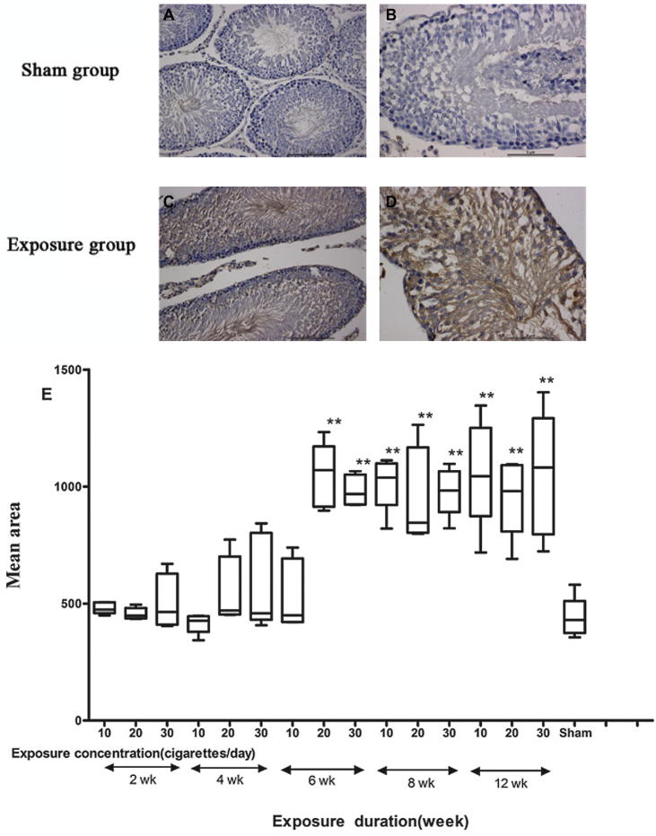
Figure 4.
Immunohistochemical analyses with caspase-3 for the detection of apoptosis. (A) and (B) Testicular tissues of sham group. (C) and (D) Testicular tissues of smoke-exposure groups. (E) Mean area, (F) mean density and (G) mean integrated optical density (IOD) of caspase-3 staining in each group. *P ⩽ 0.05, **P ⩽ 0.01 for the respective sham.


