Fig. 1.

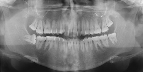
Panoramic radiograph obtained before biopsy, showing an osteolytic lesion in relation to periapex of the first upper left molar and projecting toward the floor of the maxillary sinus (white arrows)

Panoramic radiograph obtained before biopsy, showing an osteolytic lesion in relation to periapex of the first upper left molar and projecting toward the floor of the maxillary sinus (white arrows)