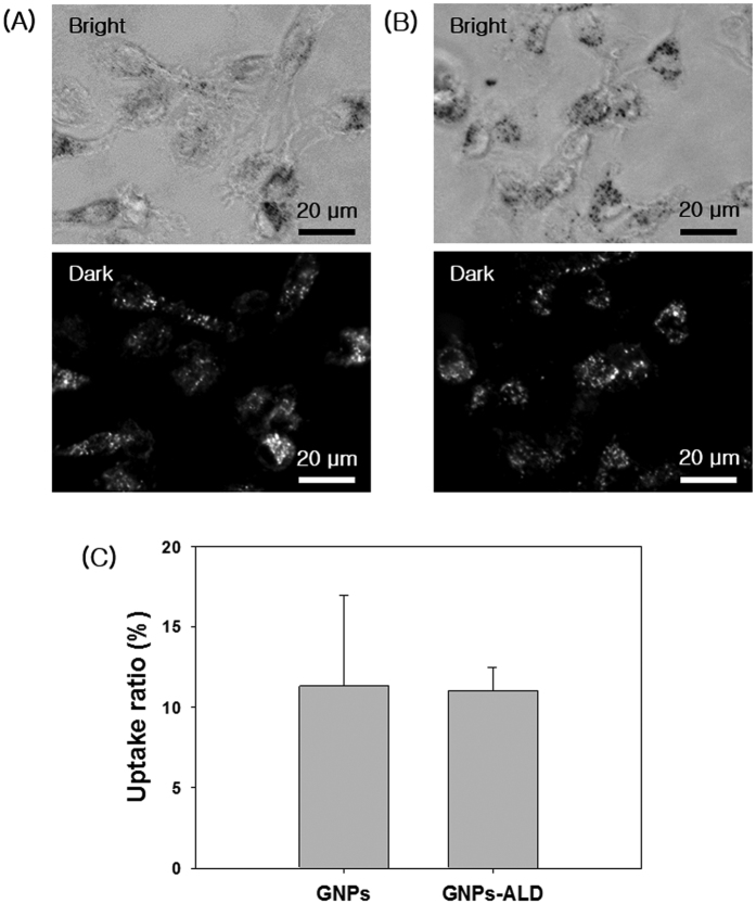
Figure 2.
Optical images and dark field images of BMMs incubated with 20 μM GNPs (A) and GNPs-ALD (B) at 37 °C, 5% CO2 for 12 h. The scattering images calculated by Image J program (bright spot area per total area), and the results are shown as mean ± SD of triplicate experiments (n = 3). Scale bars are 20 μm. Images collected at 400× magnification.

