
Fig. 5.
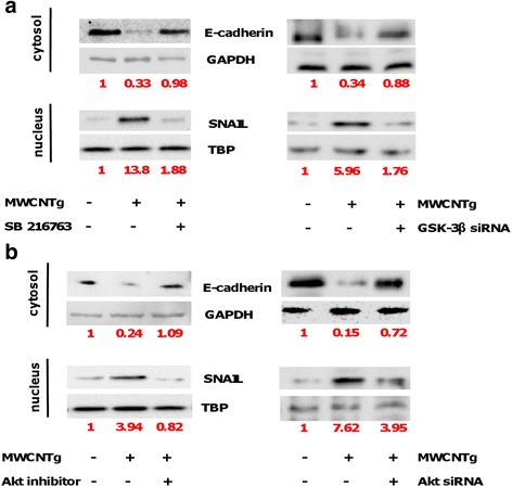
Akt and GSK-3β inhibition effect in BEAS-2B cells exposed to MWCNTg. The cells were incubated in either the absence (0 μg/ml, control) or presence of 44 μg/ml of MWCNTg for 96 h, without and with 5 μM SB216763 or 10 nM GSK-3β siRNA (a), without and with 5 μM of the specific Akt 1/2 Kinase Inhibitor or 50 nM Akt 1/2 siRNA (b). Each figure is representative of three experiments giving similar results. Results were expressed in units of relative protein expression compared to control cells (indicated in red in the figure)