
Fig. 1.
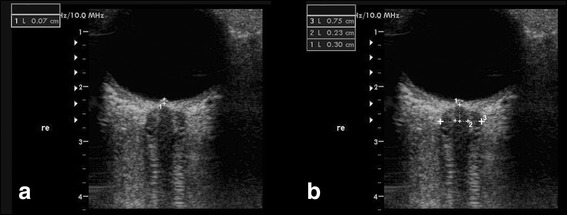
TOS in B mode of the eyeball and the optic nerve in a patient with IH. Panel a Optic disc elevation (ODE) is gauged between the fundus and the dome of the papilla in a patient with IH. Panel b Optic nerve sheath diameter (ONSD) and optic nerve diameter (OND). ONSD and OND were measured 3 mm behind the papilla (1) in an axial plane showing the optic nerve in its longitudinal course. The dotted lines denote the OND (2) and the ONSD (3)