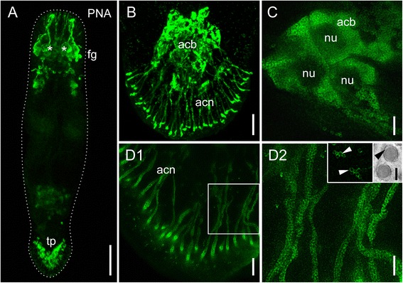
Fig. 2.

Fluorescence microscope-, confocal-, and gSTED images of PNA labelled Macrostomum lignano. a Overview of a PNA labelled adult animal. Asterisks indicate position of eyes. b Tail plate of a hatchling (confocal projection) with labelled adhesive gland cells. (c-d2) Projections of gSTED images. c Adhesive gland cell bodies with labelled vesicles in the cytoplasm, dark areas represent the nucleus. (d1) Gland cell necks filled with vesicles and (d2) detail thereof. Inset 1: detail of stained vesicles, arrowheads indicate the circular staining surrounding an unstained center. Inset 2: TEM image of adhesive gland vesicles, arrowhead indicates the lucid rim surrounding an electron-dense core. Acb adhesive gland cell bodies, acn adhesive gland cell necks, fg frontal glands, nu nucleus, ph pharyngeal glands, tp tail plate. Scale bars: (A) 100 μm, (B) 10 μm, (C,D2) 2 μm, (D1) 5 μm, (D inset) 200 nm
