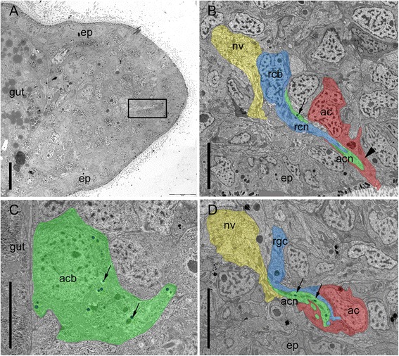
Fig. 5.

Ultrastructure of Macrostomum lignano regenerating adhesive organs after 48 h of tail plate regeneration. Anterior is to the left and dorsal to the top. a Overview of the tail plate 48 h post amputation. Rectangle indicates a regenerating adhesive organ. b Differentiated adhesive organ with anchor cell emerging through the epidermis. Arrowhead indicates intermediate filaments in the anchor cell. Arrow indicates a vesicle of the adhesive gland cell. c Adhesive gland cell body positioned at the basis of the blastema, next to the gut, with adhesive vesicles (arrows). d Immature adhesive organ, with anchor cell located beneath the epidermal layer. Note that the anchor cell is still lacking intermediate filaments and microvilli. Arrows indicate vesicles of the adhesive gland cell. Ac anchor cell, acb adhesive gland cell body, acn adhesive gland cell neck, ep epidermis, nv nerve, rcb releasing gland cell body, rcn releasing gland cell neck. Scale bars: (a) 10 μm, (b-d) 5 μm
