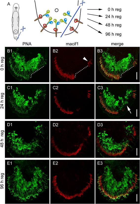
Fig. 6.

Regeneration of adhesive organs after partial amputation. a Schematic drawing of Macrostomum lignano with indication of the amputation level. The specimen were amputated on one side of the tail plate, removing the epidermal layer and the anchor cells. The cut went through the adhesive gland cell necks, but left the adhesive gland cell bodies intact. b-e PNA and Macif1 double staining of regenerating specimen at (B) 0 h, (C) 24 h, (D) 48 h and (E) 96 h after partial amputation. b The dotted line indicates the area of the cut. Arrowhead indicates anchor cells that were not affected by the cut. c After 24 h of regeneration the staining of adhesive gland cell vesicles (PNA) was reduced at the amputation side (arrow). d After 48 h the first anchor cells and adhesive gland cell necks were rebuilt. e After 96 h the regeneration of the adhesive organs was completed. Scale bars: 20 μm
