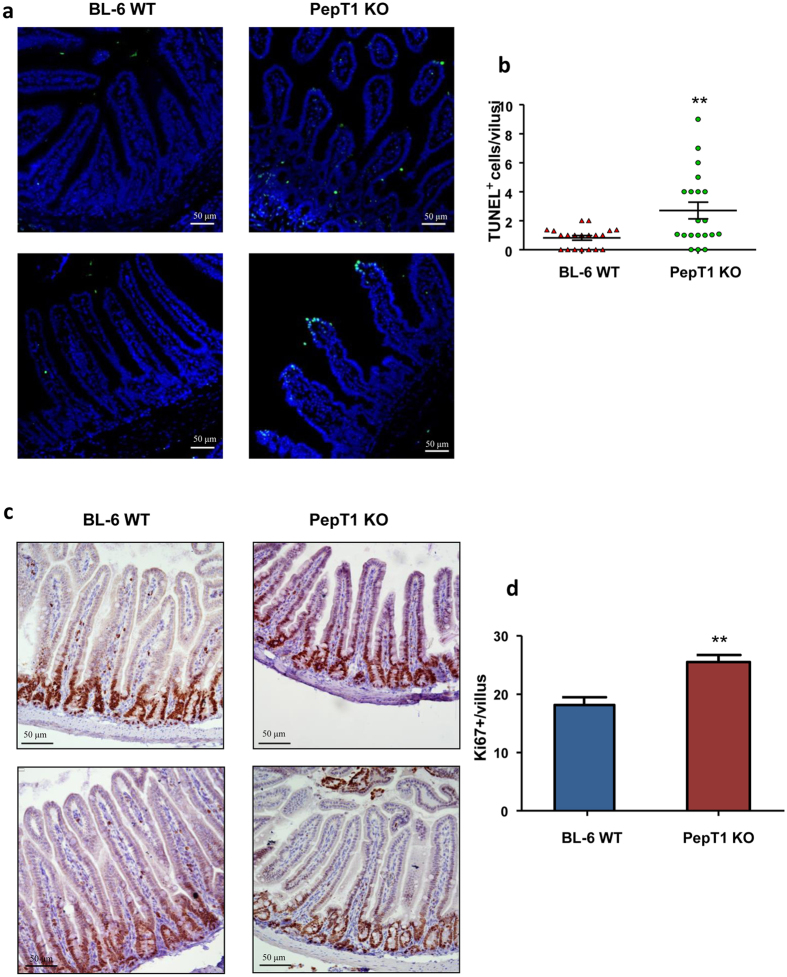
Figure 12. PepT1 KO increases small intestinal epithelial cell proliferation and apoptosis.
(a) Apoptotic small intestinal epithelial cells were quantified using a TUNEL assay (FITC, green) and nuclei were stained with DAPI (blue). (b) Cells positive for both TUNEL and DAPI staining were counted and averaged per villus. (c) The levels of epithelial cell proliferation in small intestine sections from BL-6 WT and PepT1 KO mice were assessed by immunohistochemistry using the proliferation marker, Ki67. (d) Ki67+ cells were counted and averaged per villus. Scale bars, 50 μm; **P < 0.005.

