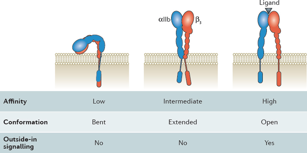
Figure 2.
Inside-out activation of αIIbβ3 platelet integrin. Drawing of the bent (left), extended (middle) and extended-open (right) conformation of αIIbβ3. αIIb in blue, β3 in red. Ligand binding site indicated by black triangle in extended-open integrin. Note the movement of the transmembrane and cytoplasmic domains with integrin activation. Binding of cytoplasmic adaptor molecules (not shown here) are thought to drive the conformational changes in the ectodomains. Ligand binding affinity, conformation and outside-in signaling noted below each conformation.

