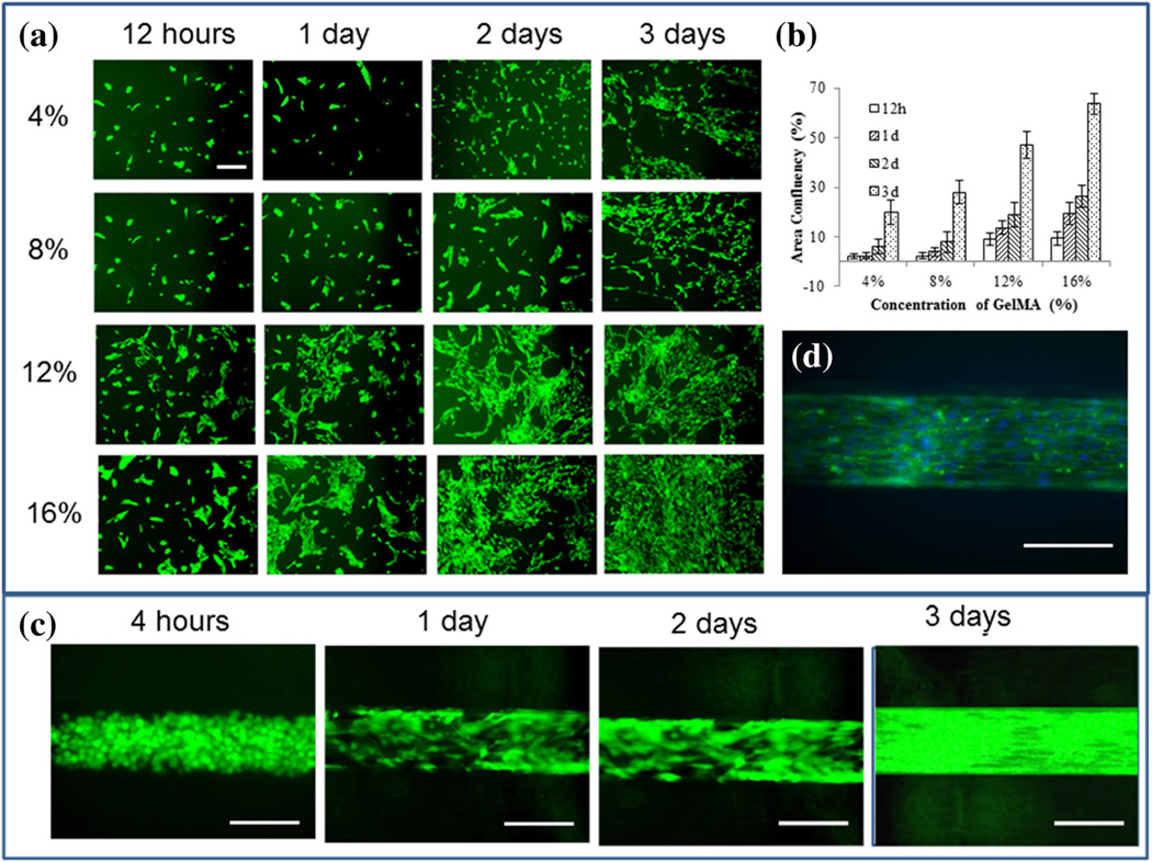
Fig. 4.
Formation of the EC monolayer on 2D GelMA surface and inside the lumen of the vascular-like structure, a GFP-fluorescent images showing attachment, spreading and growth of HUVECs on 2D GelMA surface at 4, 8, 12 and 16 % GelMA concentrations after 12 h, 1, 2 and 3 days of cell seeding, b area of confluence of HUVECs on 2D GelMA surface over time for different GelMA concentrations. c Spreading of HUVECs inside the lumen of a vascular-like structure with 12 % GelMA concentration over time, and d a representative Phalloidin-DAPI staining image showing the continuous monolayer of HUVECs in the channel at 12 % GelMA concentration after 3 days of cell seeding. The cells spread well on both the 2D surface and the luminal surface, forming a cell monolayer, as is seen from the actin skeleton (green) and the nucleus of the cells (blue). Scale bars in figures a are 100 µm while those in figures b and c are 200 µm

