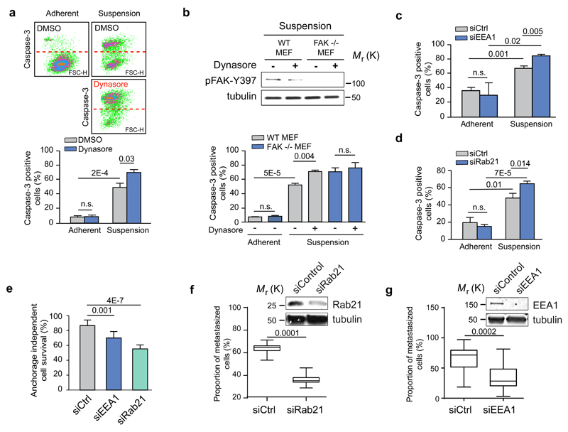
Figure 7. Integrin endosomal signalling and anoikis sensitivity are Rab21- and EEA1- dependent.
a-d, Quantification of caspase-3 positive (FL1) apoptotic cells in serum-starved TIFFs and representative dot blots (a), in FAK-/- or FAK+/+ WT MEFs ± dynasore (b) and following EEA1 (c) or Rab21 (d) silencing in TIFFs (mean fluorescence ± SEM, n=3 independent experiments). e, Quantification of anchorage-independent survival of MDA-MB-231 cells following EEA1 or Rab21 silencing (mean ± s.d., n=3 independent experiments). a-e, Student's two-tailed unpaired t-test P values are provided. f-g, MDA-MB-231 cells transfected with siCtrl and siRab21 (f) or siCtrl and siEEA1 (g) were fluorescently labelled with green or far-red cell trackers and coinjected 1:1 into the tail vein of mice. The proportion of extravasated cells was analysed by flow cytometry 48 hr after coinjection and is represented as a percentage of total extravasated cells in the lung (box plots show the 25th–75th percentiles delineated by the upper and lower limits of the box; the median is shown by the horizontal line inside the box. Whiskers indicate maxima and minima; siRab21: n = 15 mice from one experiment, siEEA1: n=10 & 15 mice pooled from two independent experiments). Student’s two-tailed unpaired t-test P values are provided. Uncropped images of blots are shown in supplementary figure 9. Statistics source data can be found in Supplementary Table 2.

