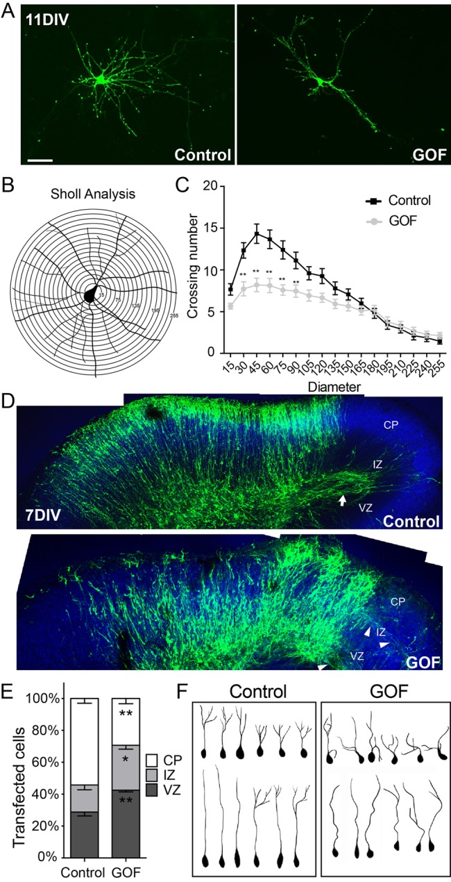
Fig. 3.

Misexpression of Sox11 in mouse cortical neurons interferes with dendritic extent in vitro and localization and shape of neurons in vivo. (A-C) Images of primary cortical neurons expressing GFP alone (left) or GFP and Sox11 (right) (A). A schematic of Sholl analysis of a cortical neuron in which neurite crossings for each ring are quantified (B). Overexpression of Sox11 (gray) reduced the complexity of dendritic branching of cortical neurons in vitro compared to control neurons (black) (C). (D) Organotypic slice cultures of cortex transfected at E14.5 with GFP alone (top) or GFP and Sox11 (bottom) and imaged at 7DIV. GFP+ cells have extended an axon tract in control (arrow) while in Sox11GOF, axons were diffuse (arrowheads). (E) Distribution of GFP+ cells in cortical embryonic zones is different in Sox11GOF compared to control; Sox11GOF cells tend to remain closer to the lateral ventricle, in the ventricular zone (VZ) and intermediate zone (IZ), than control-transfected cells. (F) Representative traces of neurons from control (left) and Sox11GOF (right) cortical plate (CP) reveals that neurons are less orderly when Sox11 expression is maintained. Scale bar: 30 μm (A); 63 μm (D). Data represented as mean±sem; *P<0.05; **P<0.01.
