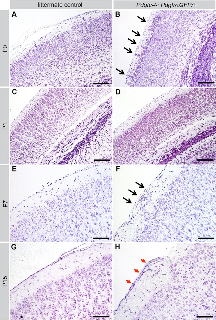
Fig. 4.
Cerebral cortex at different postnatal ages. Cresyl Violet staining of cerebral cortex of Pdgfc−/−; PdgfraGFP/+ (right) and littermate controls (left). A-F are coronal sections, G-H are sagittal. (A) Pdgfc+/−; PdgfraGFP/+ at P0. (B) Cortical neurons extend into the marginal zone (black arrows) of Pdgfc−/−; PdgfraGFP/+ at P0. (C) PdgfraGFP/+ at P1. (D) Thin cortex in Pdgfc−/−; PdgfraGFP/+ at P1. (E) Pdgfc+/−; PdgfraGFP/+ at P7. (F) Displacement of cells (black arrows) close to the meningeal border in Pdgfc+/−; PdgfraGFP/+ at P7. (G) PdgfraGFP/+ at P15. (H) Thick layer of cells outside of the marginal zone (red arrows) in Pdgfc−/−; PdgfraGFP/+ at P15. Scale bars: 150 µm.

