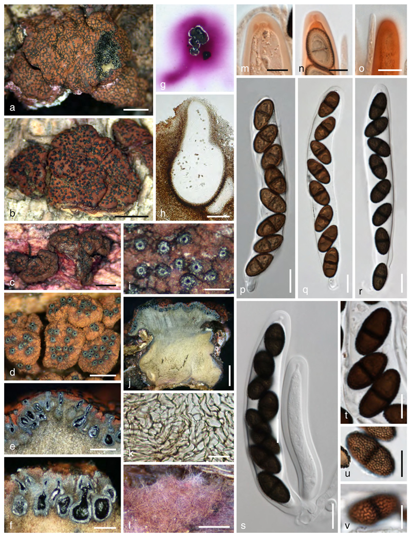
Fig. 14.
Myrmaecium rubricosum. a–d, i Ectostromata in face view (d, i. showing ostioles). e, f, j Vertical stroma sections. g Pink pigment dissolved in KOH. h Vertical section of a perithecium. k Prosenchymatous stroma tissue. l Subiculum. m–o Apical ascal rings in Congo red (note free ends of paraphyses in n and o). p–s Asci. t–v Ascospores (u, v showing surface ornamentation). Sources: a, e, j. MP133; b, g. WJ940; c. VRF; d. TH; f, m, o, t, v. VRM; h, k, n, r, s. VRP; i, l. VRJ1; p, u. isolectotype; q. WJ1247. Scale bars: a, b, j=2 mm. c, l =1 mm. d, e=0.5 mm. f=0.3 mm. h=30 μm. i=0.2 mm. k=20 μm. m, n, t–v=7 μm. o–s =10 μm

