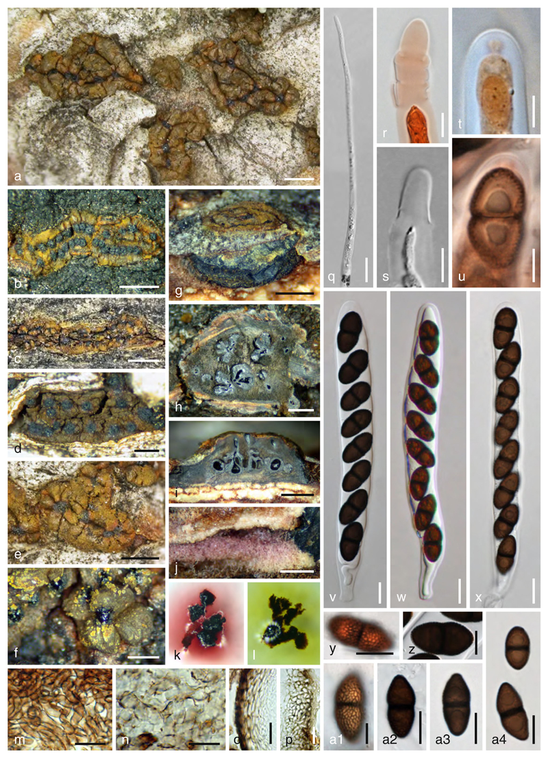
Fig. 15.
Myrmaecium fulvopruinatum. a–f Ectostromata in face view (f showing yellow pigment particles).g Stroma side view (surrounded by bark at upper level). h Transverse section at the ascomatal level. i Vertical stroma section. j Subiculum. k Purple pigment dissolved in KOH. l Yellow-green pigment dissolved in lactic acid. m, n Entostroma (m upper level; n below ascomata). k Peridium. p Section through stromatic zone. q Paraphysis. r, s Fissitunicate ascus dehiscence. t, u Apical ascal rings in Congo red. v–x. Asci. y–a4. Ascospores (y, a1. showing surface ornamentation). Sources: a, e, m–q. PWB=Picea; b, f, j. VFQ; c, v, al, a3. VFJ; d, t, u, x, a4. VFB; g, i, k, l. VF; h. VFJ1; r. WJ870; s, w. WJ1190; y. isolectotype L0819035; z, a2. VFA. Scale bars: a, d, e, h, j=0.5 mm. b, c, g, i=1 mm. f=0.3 mm. m–p=20 μm. q–s, v–z, a2-a4=10 μm. t, u, a1 = 5 μm

