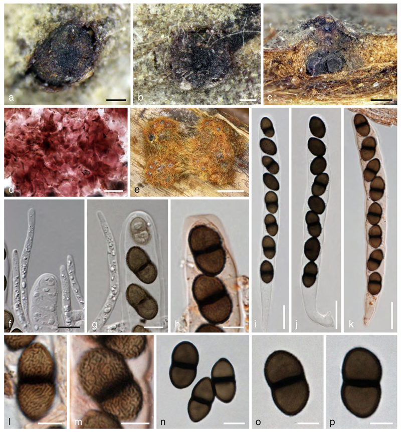
Fig. 17.
Myrmaecium rubrum (holotype). a, b Ectostromata in face view. c Stroma in side view. d Stroma tissue in KOH. e Stroma in MEA culture. f, g Free ends of paraphyses. h Ascus apex in Congo Red. i–k Asci (k. in Congo Red). l–p Ascospores (l, m showing surface ornamentation). Scale bars: a, b=0.1 mm. c=0.2mm. d, i–k=15 μm. e=1 mm. f, g, n=7 μm. h, l, m, o, p=5 μm

