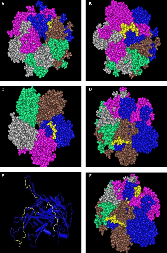
Figure 7.
Display in space-fill rendering of HPV11 L1 protein pentameric structure (2R5K). The conserved surface-exposed segments identified by comparison of protein variability and ASA profile analysis, as shown in Figure 2 and given in Table 2, are highlighted here in yellow using the protein shown in blue as a template. Peptide stretches shown are in terms of peptide stretch starting position numbers: (A) 142, (B) 264, (C) 305, and (D) 352. (E) The worm view of the protein with the four peptides highlighted in yellow. (F) Another view of the three peptides on the template in the pentameric structure.

