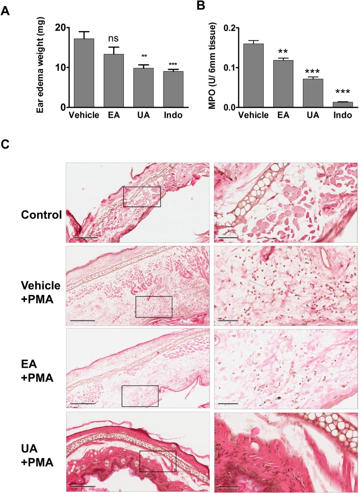
Fig 7. UA treatment reduces PMA-induced ear edema and MPO activity.
Mice (n = 5) were orally gavaged with vehicle control (10% glucose), UA (40 mg/kg body weight), EA (40 mg/kg body weight) and indomethacin (10 mg/kg body weight) at 2 hrs prior to applying PMA to right ear and acetone (control) to the left ear. At six hrs post application of PMA, animals were sacrificed by cervical dislocation and (A) weight of edema and (B) MPO activity was evaluated as described in material and method section. Results are expressed as mean ± SEM. *, ** and *** indicates p value < 0.05, 0.01 and 0.001, respectively compared to vehicle group. (C) Representative H&E images of ear edema at 100x magnification (left) and 400x magnification (right) indicate decreased PMA induced inflammation i.e., infiltration of inflammatory cells in UA treated animals compared to vehicle or EA treated animals. The images were captured using Aperio Imagescope and scale bars on 100x and 400x magnifications indicate 300 μm and 60 μm respectively.

