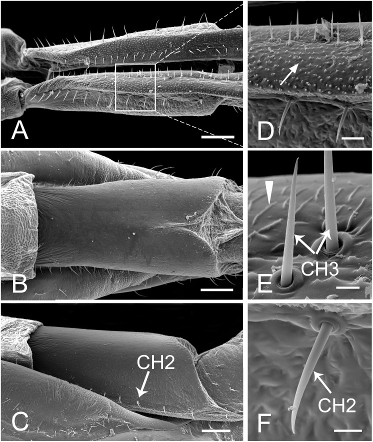
Fig 3. SEM of first segment of labium of female Lycorma delicatula.
(A) Anterior view. (B) Ventral view. (C) Lateral view. (D) Enlarged view of outlined box of (A), showing the papillae (white arrow). (E) Enlarged view of sensilla chaetica III (CH3) and prominent transverse ridge (white triangle). (F) Enlarged view of sensilla chaetica II (CH2). Bars: (A), (B) and (C) = 150 μm; (D) = 30 μm; (E) = 6 μm; (F) = 12 μm.

