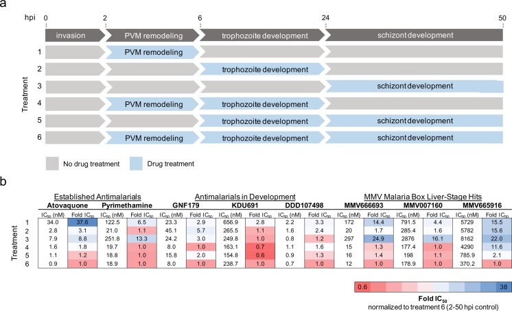
Figure 7.
Exoerythrocytic-stage active compounds display unique potencies during exoerythrocytic-stage EEF development. (a) Diagram illustrating the major stages of malaria parasite exoerythrocytic-stage development, including invasion, parasitophorous vacuole membrane (PVM) remodeling, trophozoite development, and EEF schizont development. During the first 4 h after sporozoite invasion, the parasite dramatically remodels its parasitophorous vacuole membrane by degrading host-cell-derived proteins and at the same time inserting its own parasite-derived proteins.6 During the next 18 h, the sporozoites transform from their elongated motile form to round, nonmotile, and metabolically active trophozoites. The trophozoites undergo impressive nuclear replication starting at around 24 h postinfection, displaying one of the fastest replication rates known to eukaryotic organisms to develop into mature EEFs.6 Drug treatments 1–6, corresponding to compound incubation during the exoerythrocytic developmental stages indicated, are shown. (b) Pb-Luc IC50 data for established antimalarial compounds (atovaquone and pyrimethamine), antimalarials in development (GNF179, KDU691, DDD107498), and the three MMV Malaria Box compounds (MMV666693, MMV007160, and MMV665916) added during Pb-Luc exoerythrocytic-stage development in a modified 384-well luciferase-based assay (discussed in Materials and Methods) are shown. Likewise, the Pb-Luc IC50 fold changes normalized to the 2–50 h drug-treated controls are shown and colored based on the indicated heat map.

