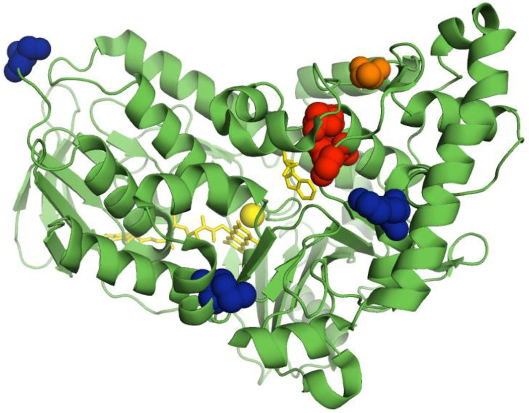
Figure 1.
Location of mutations in RebH variants employed in this work. A previously reported crystal structure of wild-type RebH (PDB entry 2OA1)23 with residues that are mutated in variant 1-PVM shown in blue, additional residues mutated in variant 3-SS shown in red, and residue A442 further mutated in variant 4-V shown in orange.22 Bound L-tryptophan, FAD, and chloride are shown in yellow.

