
Figure 2. BI 2536 and VCR cooperate to induce caspase-dependent apoptosis.
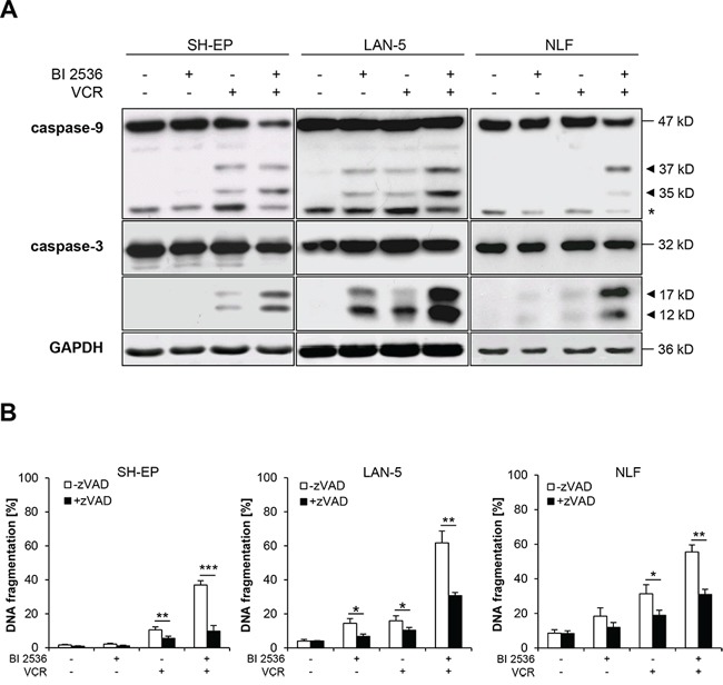
A. SH-EP cells were treated with 7.5 nM BI 2536 and/or 5 nM VCR, LAN-5 cells with 30 nM BI 2536 and/or 30 nM VCR, NLF cells with 4 nM BI 2536 and/or 0.5 nM VCR for 24 hours. Activation of caspase-9 and -3 was analyzed by Western blotting. Arrowheads indicate active cleavage fragments, expression of GAPDH served as loading control, asterisk denotes unspecified bands. Representative blots of two independent experiments are shown. B. SH-EP, LAN-5 and NLF cells were treated for 48 hours as described in (A) in the presence or absence of 20–50 μM zVAD.fmk. Apoptosis was determined by analysis of DNA fragmentation of PI-stained nuclei using flow cytometry. Data are shown as mean +/− SD of three independent experiments performed in triplicate; *, P < 0.05; **, P < 0.01; ***, P < 0.001.