
Figure 3. Cr(VI) induces the DNA damage response.
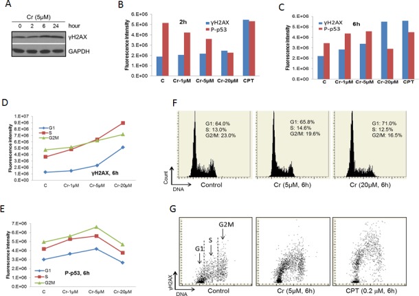
A. BEAS-2B cells were treated with 5 μM Cr(VI) for the indicated time periods. At the end of the treatments, total proteins were extracted from the cells and subjected to Western blot with antibodies against the indicated proteins. B. & C. BEAS-2B cells on chamber slides were treated with Cr(VI) at the indicated concentrations for 2 hours B. or 6 hours C. followed by LSC as described in Materials and Methods. The total levels of γH2AX and P-p53 were plotted against the concentration of Cr(VI). D. & E. Data from B and C were analyzed by splitting the cells in different phases of the cell cycle according to the DNA content. The levels of γH2AX D. or P-p53 E. in cells of different phases of the cell cycle were plotted against the concentration of Cr(VI). F. Examples of the readout of the cell cycle analysis showing percentages of cell in different phases of the cell cycle. G. Examples of the readout of the γH2AX measurement.