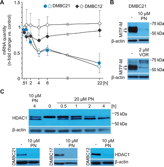
Figure 4. MITF level in melanoma cells might be reduced via inhibition of HDAC1 activity.

A. Expression of total MITF (closed symbols) and MITF-M (open symbols) was analyzed by qRT-PCR in DMBC21 and DMBC12 melanoma cell populations treated with 20 μM PN. n-fold change in mRNA quantity is represented after normalization to RPS17 and the respective DMSO-treated control. B. Immunoblot analysis of lysates from DMBC21 cells treated with either 10 μM PN or 2 μM vorinostat (VOR) for 24 hours. C. DMBC21 cells were treated with 10 μM and 20 μM PN and harvested for Western blots at different time points to show changes in the HDAC1 level (top). HDAC1 level was assessed after 24 hours incubation with 10 μM PN (bottom). In Western blot experiments, equal loading was confirmed by β-actin. Representative results are shown.
