
Figure 5. OAd-infected MSCs invade tumors in vivo.
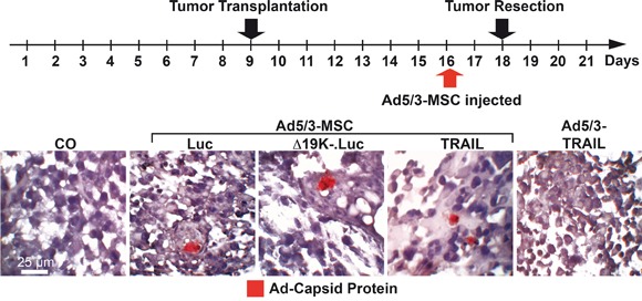
At developmental day 9, 5×10⁵ MIA-PaCa2 cells were transplanted to the CAM of chick embryos (18 eggs per group). At day 16, the CAM was injected with 100 μL of cell culture medium (CO), 1×10⁸ TCID50 Ad5/3-TRAIL in 100 μL of cell culture medium, or 5×10⁴ MSCs in 100 μL of cell culture medium 2 h after infection with 2000 TCID50 of Ad5/3-Luc (Luc), Ad5/3-Δ19K-.Luc (Δ19K-) or Ad5/3-TRAIL (TRAIL). The tumor xenografts were resected at day 18. Tissue sections were prepared and paraffin-embedded, and the expression of adenoviral capsid protein (red) was detected by immunohistochemistry. Representative images at 400× magnification are shown.