
Figure 6. MSC-delivered OAds reduce tumor growth in vivo.
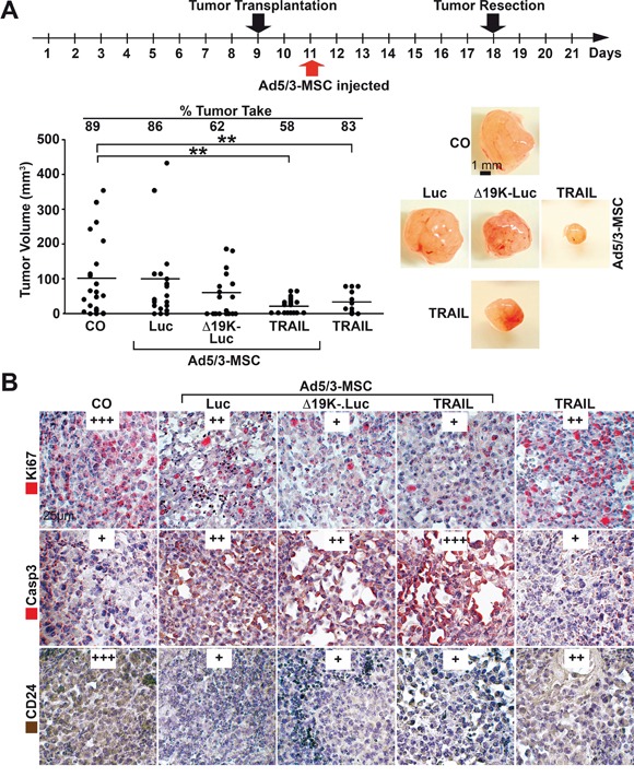
A. Experimental procedure: MIA-PaCa2 cells (5×10⁵) were transplanted to the CAM of chick embryos at developmental day 9 (20 eggs per group), followed by therapeutic injection at day 11, as described in Figure 5, and tumor resection at day 18. The tumor take rate and the volume of the resected xenografts were determined, and the single data points and the means of each group are shown (**P<0.01) along with representative images of tumor xenografts of each group. B. Sections were prepared from the xenograft tumors and paraffin-embedded, and the expression of Ki67, cleaved fragment of Caspase 3 (Casp3) and CD24 were examined by immunohistochemistry. Representative images are shown at 400× magnification. The expression levels were evaluated by a semi-quantitative scoring system as described in Figure 4B.