
Figure 3. Immunohistochemical expression of CXCR4 and somatostatin receptors 2a and 5 in SCLC.
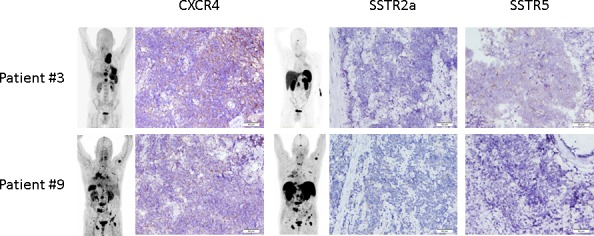
Display of two examples of immunohistochemical expression of CXCR4 and SSTR2a and 5, respectively. Patient #3 had his biopsy taken from a lymph node metastasis demonstrating a weak staining for CXCR4 in 90% of the tumor cells (IRS 4). SSTR2a was negative, SSTR5 could also be demonstrated to be weakly expressed in 90% (IRS 4). Patient #9 also presented with extensive disease. Biopsy of the primary tumor revealed mild CXCR4 (intensity 1+ in 70% of the cells, IRS 3) and mild SSTR2a (intensity 1+ in 90%; IRS 4) expression. SSTR5 was negative in the sample. The inserts depict maximum intensity projections are the respective whole-body [68Ga]Pentixafor- and SSTR-directed PET/CT scans, respectively.