
Figure 4. Evaluation of endogenous GTPCH expression in human breast tissue-derived fibroblasts on breast cancer cell proliferation.
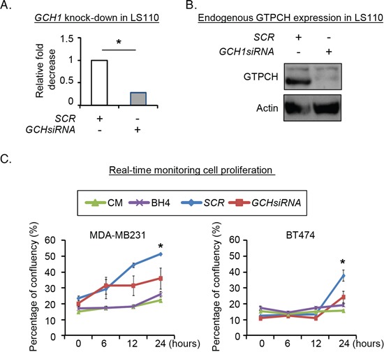
Human breast fibroblast LS110 was transfected with GCH siRNA or SCR siRNA, respectively for 48hours. RNA from the cells were extracted and quantified for GCH1 mRNA (A). Cell lysates were prepared for immunoblotting analysis with antibodies to GTPCH or GAPDH (representative 3 independent experiments). (B). 2 × 10⁴ of MDA-MB231 or BT474 cells/well were seeded in 12-well plates and treated with conditioned media (CM) of GCHsiRNA or SCRsiRNA transfectants, or CM ± BH4 (100μM). Images were taken and analyzed by real-time Incucyte for 24 hours (C). All data are shown as mean ± SEM (*p <0.05 vs. SCR control, n = 3).