
Figure 2. AC-73 disrupts CD147 dimerization.
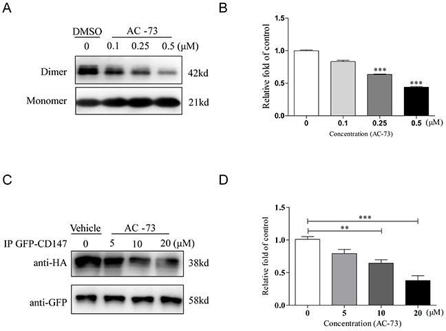
A. Representative image of CD147 dimerization. A total of 5 μg of purified CD147 was mixed with 5× Laemmli sample buffer lacking SDS and different concentrations of AC-73 (0.1, 0.25, or 0.5 μM). DMSO was used as negative control. The material was then resolved on a 10% SDS-PAGE gel without boiling, followed by immunoblotting with an anti-His6 antibody. The dimer bands were approximately 42 kDa in size. B. Quantification of CD147 dimerization inhibition by densitometry analysis. C. AC-73 inhibited CD147 dimerization in 293T cells, as determined using a co-IP assay. D. Quantification of CD147 dimerization inhibition in cellulo by densitometry analysis. The bars represent the mean of triplicate measurements of each sample, and the error bars indicate ± SD. ***P < 0.001, **P < 0.01, *P < 0.05, one-way ANOVA (H).