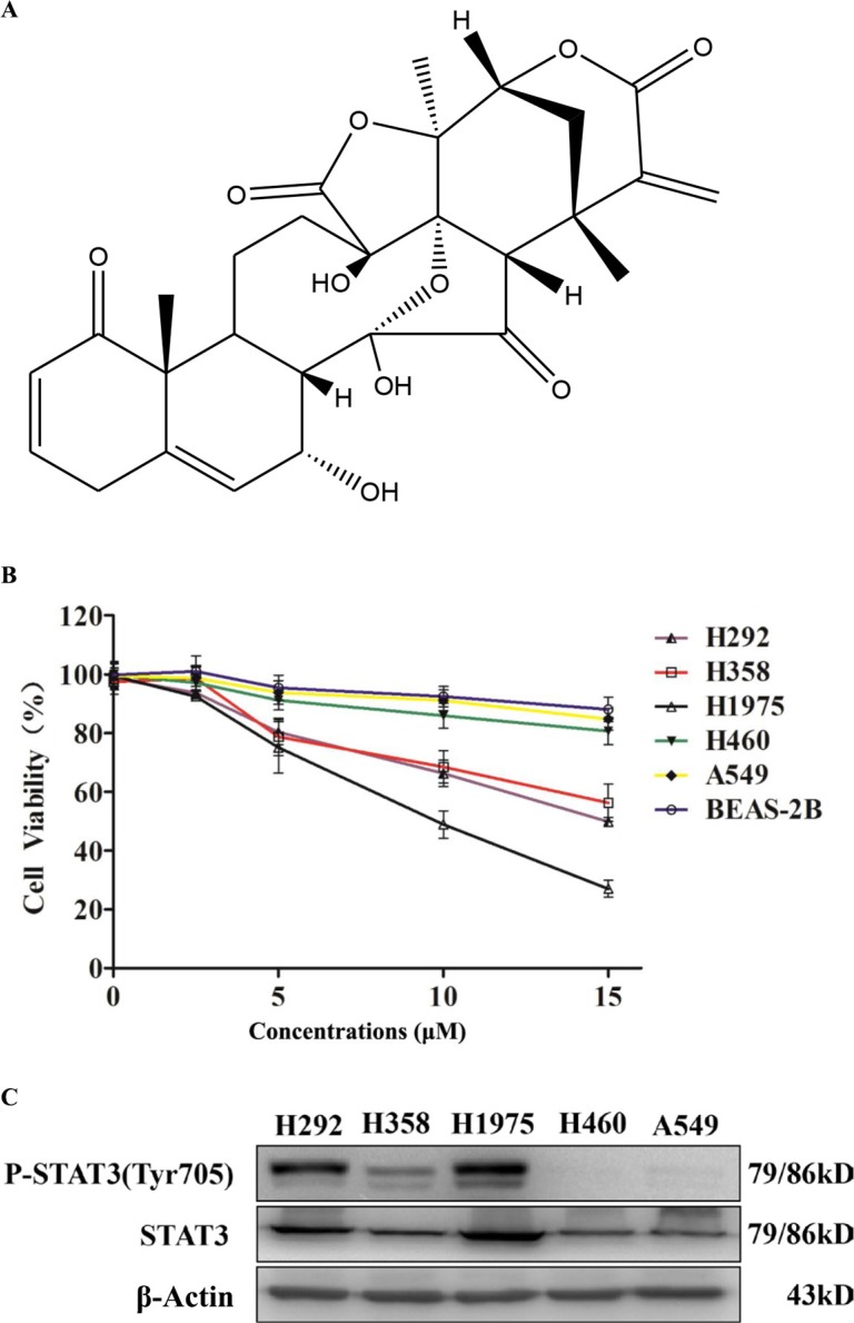
Figure 1. Physalin A exerts anti-proliferative effects in human NSCLC cells with activated STAT3.
(A) Structure of physalin A. (B) The human NSCLC cell lines, H292, H358, H1975, H460, A549, and BEAS-2B (1 × 10⁴ cells/well) were treated with the indicated concentrations of physalin A for 24 h. Cell viability was then measured using the CCK-8 assay. Results are presented as mean ± SD from three independent experiments. (CB) p-STAT3 (Tyr 705) and STAT3 levels were detected in the H292, H358, H1975, H460 and A549 cell lines. β-actin was used as a loading control.

