
Figure 5.
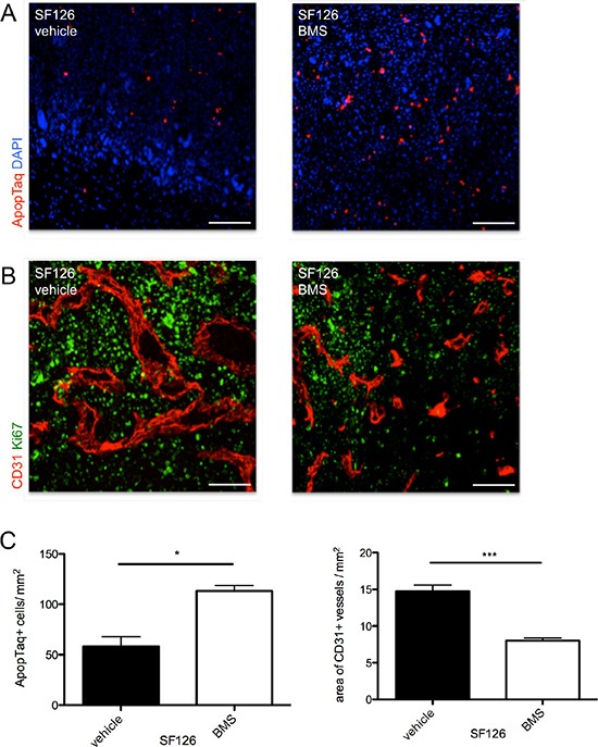
(A) Intratumoral apoptotic events in SF126 xenografts under treatment with BMS-777607 and vehicle. (B) CD31 staining (red) shows differences in vessel size and density under treatment with BMS-777607 in vivo. Proliferative activity is displayed qualitatively with Ki67 staining (green). (C) Images show corresponding statistical analysis of apoptotic events within tumor tissue (n = 3, *p = 0.0121) and statistical analysis of intratumoral vessel density (n = 3, ***p < 0.0001). Scale bar indicates 50 μm.